Anatomy Muscles Pelvis ~ The Pelvic Floor Muscles Part 1 Basic Anatomy Youtube. The muscles of the pelvis, hip and buttock anatomical chart shows how each muscle in this area of the body works with the others, and the various minor systems within the major ones. Muscles that attach from the pelvis to the trunk and cross the lumbosacral joint muscles that attach from the pelvis to the thigh/leg and cross the hip joint pelvic floor muscles that are located wholly within the pelvis Attached to the pelvis are muscles of the buttocks, the lower back, and the thighs. Anatomy muscles pelvis / pelvis muscles diagram function body maps : The muscles of the pelvis form its floor.
The thigh bone or femur and the pelvis join to form the hip joint. (2) the levator ani and the coccygeus, which together form the pelvic diaphragm and are associated with the pelvic viscera. Rectus femoris muscle, one of the quadriceps muscles on the front of your thigh. The levator ani muscles consist of three. The floor of the pelvis is made up of the muscles of the pelvis, which support its.
Pelvis Anatomy Muscles Anatomy Drawing Diagram from i.pinimg.com On the posterior side they are the glutei and on the anterior side the hip muscles extending into the thighs. These muscles have attachments to the pelvis as follows: The piriformis is a triangular muscle 1 on either side on the very front of the posterior wall of true pelvis. Several muscles around the pelvis take part in movements of the thigh. Pelvis anatomy muscle thigh muscular system pelvis text hand human png pngwing from w7.pngwing.com this mri pelvis cross sectional anatomy tool is absolutely free to use. The psoas major and iliacus make up the iliopsoas group. Learn about anatomy muscles pelvis with free interactive flashcards. Most muscles that insert on the femur (the thigh bone) and move it, originate on the pelvic girdle.
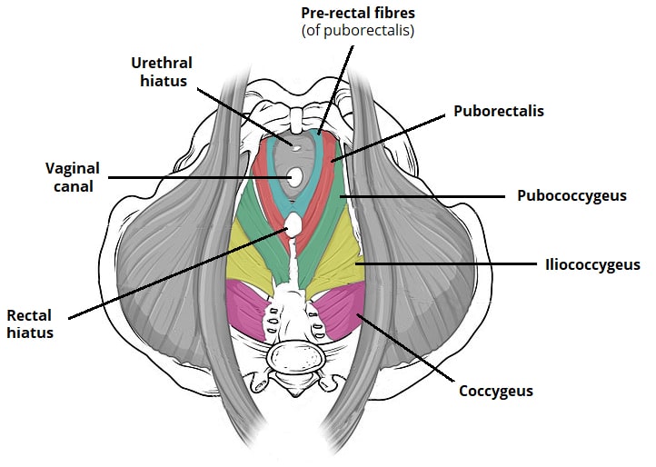
The levator ani is a broad sheet of muscle.
They are also known as the inner hip muscles and deep external rotators. The main function of the pelvic floor muscles are: Anatomy muscles pelvis / pelvis muscles diagram function body maps : Muscles an important group of muscles in the pelvis is the pelvic floor. The classification of the two groups under a common heading is. Some of the major pelvic muscles are as follows. The levator ani muscles are the largest group of muscles in the pelvis. Rectus femoris muscle, one of the quadriceps muscles on the front of your thigh. Included in this group are the adductor longus, adductor brevis, adductor magnus, pectineus, and gracilis muscles. Here we explain the hip and groin muscles, their actions and exercises. The muscles of the pelvis form its floor. Large ligaments, tendons, and muscles around the hip joint hold the bones (ball and socket) in place and keep it from dislocating. Anatomy pelvis muscles muscles of the pelvis.
They are also known as the inner hip muscles and deep external rotators. Included in this group are the adductor longus, adductor brevis, adductor magnus, pectineus, and gracilis muscles. The levator ani muscles consist of three. The muscles of the pelvis, hip and buttock anatomical chart shows how each muscle in this area of the body works with the others, and the various minor systems within the major ones. These muscles move the thigh toward the body's midline.
Pelvis Anatomy Muscles Anatomy Drawing Diagram from i.pinimg.com Included in this group are the adductor longus, adductor brevis, adductor magnus, pectineus, and gracilis muscles. These muscles move the thigh toward the body's midline. Several muscles around the pelvis take part in movements of the thigh. The gluteal muscles are a group of three muscles named the gluteus maximus, the gluteus medius, and the gluteus minimus. These muscles have attachments to the pelvis as follows: There are around 650 skeletal muscles within the typical human body. The small intestine is the longest part of the. Arcus tendineus levator ani and the ischial spine
The muscles within the pelvis may be divided into two groups:
Anatomy of pelvic and acetabular muscles. They have several functions, including helping to support the pelvic organs. Large ligaments, tendons, and muscles around the hip joint hold the bones (ball and socket) in place and keep it from dislocating. The thigh bone or femur and the pelvis join to form the hip joint. The pubococcygeus (pc) muscle is the muscle that runs the show in pelvic floor health. The classification of the two groups under a common heading is. The muscles of the pelvis form its floor. Hip muscles the hip muscles include pelvic and groin muscles. They are important for stabilising the body and for moving the legs. Most muscles that insert on the femur (the thigh bone) and move it, originate on the pelvic girdle. The pelvis is the lower portion of the trunk, located between the abdomen and the lower limbs. Some of the most important include the major digestive organs, the intestines. The adductor muscle group, also known as the groin muscles, is a group located on the medial side of the thigh.
Several muscles around the pelvis take part in movements of the thigh. The pelvic floor muscles provide foundational support for the intestines and bladder. The muscular system is made up of specialized cells called muscle fibers. Some of the largest and most powerful muscles in the body are the gluteal muscles or gluteal group. The muscles of the pelvis, hip and buttock anatomical chart shows how each muscle in this area of the body works with the others, and the various minor systems within the major ones.
The Pelvic Floor Structure Function Muscles Teachmeanatomy from teachmeanatomy.info Muscles play an important role in the. Arcus tendineus levator ani and the ischial spine The levator ani muscle has a linear origin from the pelvic outermost layer of the body of pubis, a tendinous arch of obturator fascia, and the. The muscles of the pelvis form its floor. Small and deep muscles which mainly externally rotate the thigh at the hip joint and stabilize the pelvis. They are important for stabilising the body and for moving the legs. There are around 650 skeletal muscles within the typical human body. Muscles that attach from the pelvis to the trunk and cross the lumbosacral joint muscles that attach from the pelvis to the thigh/leg and cross the hip joint pelvic floor muscles that are located wholly within the pelvis
They are important for stabilising the body and for moving the legs.
Muscles an important group of muscles in the pelvis is the pelvic floor. The small intestine is the longest part of the. This is a table of skeletal muscles of the human anatomy. Arcus tendineus levator ani and the ischial spine There are around 650 skeletal muscles within the typical human body. The pelvic floor muscles provide foundational support for the intestines and bladder. These muscles have attachments to the pelvis as follows: Muscles that attach from the pelvis to the trunk and cross the lumbosacral joint muscles that attach from the pelvis to the thigh/leg and cross the hip joint pelvic floor muscles that are located wholly within the pelvis They are important for stabilising the body and for moving the legs. The pelvis is the lower portion of the trunk, located between the abdomen and the lower limbs. The adductor muscle group, also known as the groin muscles, is a group located on the medial side of the thigh. Arcus tendineus levator ani and the ischial spine It's supplied by ventral rami of first and 2nd sacral nerves (s1, s2).
Berbagi :
Posting Komentar
untuk "Anatomy Muscles Pelvis ~ The Pelvic Floor Muscles Part 1 Basic Anatomy Youtube"


Posting Komentar untuk "Anatomy Muscles Pelvis ~ The Pelvic Floor Muscles Part 1 Basic Anatomy Youtube"